The best (and most well-paid) pitchers in baseball tend to be those with an excellent command of the curveball, able to throw at high velocity while also ensuring that the ball traces the arc of a curve with the utmost precision. Scleral lens design also relies on precise control of the curve.
As in baseball, success stems from a solid foundation in the principles of physics. But scleral lens design is quite a bit simpler—just a matter of adjusting curves and manipulating distances to achieve the desired effects of sagittal depth. This article provides an overview of key facts that can help you master scleral contact lens fitting
Scleral Design 101
There are three main components to scleral lens design: the optic zone, the transition zone over the limbus and the landing zone on the sclera. Each zone is comprised of one or more curvatures, each of which has defined diameters. The shape of the curvatures may vary; they can be spherical, aspheric or a spline (a smooth polynomial function that is more complex than a simple asphere). Each of these zones has a different function and relationship to the ocular surface.
The optic zone is the power center of the lens; it is designed to vault the cornea and protect its optical function. The transition zone raises and lowers the optical zone relative to the eye, and is also vital to protecting the limbal stem cells. The landing zone is the area in which contact between the lens and ocular surface is made. To prevent inflammation, this contact must be done in a very controlled manner.

|
|
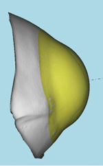
Fig. 1. The profile and sagittal depth of a standard geometry 16.1mm lens.
|
Scleral lenses are designed to vault both the cornea and limbus entirely and to land solely on the sclera. As such, it is important to understand the ways in which each zone can be manipulated to achieve the desired effect. It really is as simple as asking yourself, “Do I have too much/too little sagittal depth? What options do I have to change it?”

|
|
Fig. 2. A 0.5mm increase in transition zone curve width increases sagittal depth nearly 900 microns.
|
Small-diameter corneal GP lenses are designed to align to the corneal curvature. In these designs, the base curve (BC) has the greatest effect on vault and fluorescein patterns. Scleral lenses differ from these smaller-diameter lenses in that the transition zone is the most important zone for vault determination, followed by the overall diameter.
In scleral designs, the BC, or the back surface optic zone curvature, should be thought of foremost as a power-defining curve, and not entirely as a fit curve. Depending on which transition curves are selected, the BC may be flat or steep—affecting the power of the lens. For example, a -10.00D lens with a BC of 55.00D would be plano with a BC of 45.00D; depending on which transition zone curves are selected in this scenario, each of these BC values may have the same sagittal depth.

|
|
Fig. 3. A 1.8mm change in BC is needed to achieve the same increase in sagittal depth.
|
Steeper and wider transition curves offer greater sagittal depth, while bigger lenses allow for wider curves. Figure 1 shows the profile and sagittal depth of a standard geometry 16.1 scleral lens. By increasing the width of the transition zone curves (noted by the red boxes) by 0.5mm the sagittal depth of this lens increases nearly 800 microns (Figure 2). Conversely, a 1.8mm change in BC is needed to achieve the same increase in sagittal depth (Figure 3).

|
|
Fig. 4. The secondary curve system was steepened by 1.9mm to gain 800 microns of sagittal depth increase.
|
A reverse curve in the transition zone (i.e., the secondary curve is steeper than the base curve) can also significantly increase sagittal depth—if a change in diameter is not desired (Figure 4). In this example, the secondary curve system was steepened by 1.9mm to reach the 800 microns of sagittal depth increase.
Fitting Considerations
There are a number of ocular surface conditions that affect lens design to consider, such as corneal geometry, corneal diameter, limbal geometry and scleral geometry. Lens design is simply a matter of determining the best way to align or vault these different geometries.

| |
|
Fig. 5. Different ocular geometries require different lens designs.
|
For example, post-surgical eyes with elevated grafts will need longer transition curves (Figure 5). Conversely, flat, drumhead grafts will need short—but steep—transition curves (Figure 6).
 |
 |
|
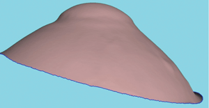
Fig. 6. Altering transition curves and widths can yield dramatic fit changes.
| |
The images on the left show the profile of a proud graft with a standard geometry scleral lens. Note the apical touch with transition zone bubble. The fit is improved by widening the transition zone curves, which increases the overall diameter. The figures on the right show a flat drumhead graft; by flattening the BC and making the transition zone curves shorter, the lens is brought closer to the cornea. Steepening the transition curves will help alleviate some of the pressure over the graft/host junction.
Focal changes (e.g., those as a result of keratoconus) will create different sagittal depths across the optic zone (Figure 7). For patients with centrally elevated corneas, a steep BC is appropriate, while a flat BC with a steep transition zone (reverse curve) will fit periphery elevations (e.g., pellucid marginal degeneration) more evenly.
|
|
|
Fig. 7. Keratoconus patients will have different sagittal depths across the optic zone.
|
Bear in mind that the limbus itself may vary in geometry along different parts of the eye. While the majority of normal eyes will be tangentially aligned from the peripheral cornea to the sclera (Figure 8, left side), some limbal areas will have severe curvature changes—as a result of either pathology or surgery (Figure 8, right side).
 |
| Fig. 8. Variations in the geometry of the limbus along different parts of the eye. While the left side of the limbus is aligned from the peripheral cornea to the sclera, note the severe curvature changes due to surgery on the right side of the limbus. |
It is necessary to bathe the limbus in fluid to protect the stem cells. Any contact with the limbus may result in inflammation and/or neovascularization of the cornea. If it is necessary to increase sagittal depth over the limbus, the transition zone curves should be widened; shortening the curves will bring the lens closer to the limbus.
The landing zone of the lens should align with the sclera while avoiding localized impingement of both conjunctival and scleral vessels. The narrower the landing zone width, the less area the lens has to land and “float” on the scleral lens surface. If the landing zone is too narrow, the lens may “sink” into the sclera, causing an impression ring (Figure 9). If you notice that the lens has an impression ring or rebound hyperemia upon removal, flatter and wider landing zone curves should be ordered.
|
|
|
Fig. 9. An impression ring due to the lens "sinking" into the sclera.
|
Changes in scleral landing zone geometry can be frustrating, with localized changes (e.g., pinguecula and pterygium) causing inflammation and discomfort. Even eyes with no scleral pathology present may have significant changes in elevation (Figure 10).
 |
| Fig. 10. Elevation changes can occur in both diseased and healthy eyes, as shown in this map of a patient with no scleral pathology. |
The nasal quadrant is typically higher and flatter in elevation than the temporal quadrant, resulting in impingement of nasal vessels. For the ultimate scleral design, bitoric, quadrant-specific or independent elevation (patient specific periphery) lenses can be ordered.
Play Ball
Scleral lens design simply comes down to whether a specific area of the lens needs to be closer to or farther away from the ocular surface. Changing curvature widths will have the biggest impact on increasing or decreasing sagittal depth. Using the transition zone to raise and lower the lens allows for flatter base curves and less minus powers.
The scleral landing zone, which accounts for focal or quadrant scleral geometry changes, can be manipulated independently from the base curve and transition zone. Wider landing zones prevent the lens from “sinking” into the sclera, so care should be taken that widening the transition curves does not come at the expense of the landing zone width.
Remember: for scleral lenses and baseballs alike, it’s simply a matter of knowing the simple principles of physics—and you have more control than you think.

