 From fungal keratitis to an update on the 2006 Fusarium outbreak, we have recently covered several infectious diseases. Now, it is time to revisit another past culprit—Acanthamoeba keratitis (AK), a rare and sight-threatening disease caused by free-living protozoans. In 2007, the CDC released several public health warnings regarding an outbreak thought to be associated with the use of Complete MoisturePlus (Abbott Medical Optics) solution.1 AMO voluntarily recalled the product.
Putting this dangerous pathogen under the public’s microscope was, and continues to be, extremely important. A plethora of literature associating AK with contact lens wear has been published. Much of this literature also details the importance of hygiene and compliance, as well as analyzing solutions to determine their efficacy in killing Acanthamoeba cysts.2
While AK is most commonly associated with contact lens wear, everyone is potentially at risk for infection. It is important for practitioners to suspect AK when examining contact lens and non-contact lens wearing patients who present with telling symptoms of the disease. Increasing evidence tying Acanthamoeba to water sources means that all patients and clinicians should be knowledgeable about this dangerous pathogen.3
The Disease
Acanthamoeba is one of the most prevalent and abundant protozoa on earth and has been isolated from treated water, seawater, tap water, soil, dust, and air. The species’ life cycle consists of trophozoite and cyst stages, and if AK is not caught and treated early, may have sight-threatening consequences.
The correlation between AK and contact lens use is high: In the United States, 85% of cases occur in contact lens wearers.4 However, the disease is still quite rare—the incidence of disease is estimated at approximately one to two cases per million contact lens users.5,6 Contact lens wearers are frequently touching their eyes, therefore putting themselves at a greater risk for infection; contaminated lenses have been known to act as “vectors” for Acanthamoeba species.5
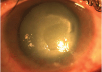
A severe case of Acanthamoeba keratitis; notice the ring infiltrate with a central epithelial defect. In cases such as this, a therapeutic corneal transplant may be necessary.
Educate Your Patient
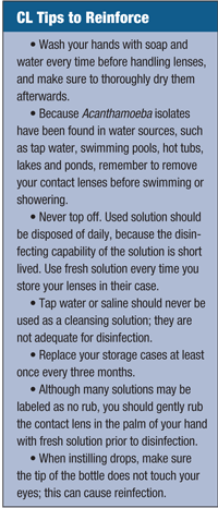
It is the practitioner’s responsibility to ensure that patients who wear contact lenses maintain a healthy and safe care regimen. Fostering open communication with your patient, as well as providing key information on compliance, is crucial in the hopes of avoiding infection (see “CL Tips to Reinforce”).
Anyone is at Risk
With the brunt of educational materials focusing on the link between AK and soft contact lens wear, practitioners may be slower to diagnose Acanthamoeba in patients with hard or rigid gas-permeable lenses, or in patients without lenses. Similarly, patients who practice appropriate lens care and hygiene practices are not completely immune to AK. However, because of its broad association with contact lens use and similar appearance to other viral, fungal and bacterial infections such as herpes simplex keratitis, AK is easy to misdiagnose. Most often, an accurate diagnosis is made when all treatments fail, or when the parasite is identified through cultures or pathological evaluation.
In a retrospective review of all AK cases diagnosed at Massachusetts Eye and Ear Infirmary between 1990 to 1994, Emil Chynn, M.D., and colleagues sought to identify potential differences in both time to diagnosis and final visual outcome between contact lens and non-contact lens users with AK.7 Results showed that mean time to diagnosis was significantly longer in non-contact lens wearing patients; 50% of non-contact lens wearing patients had a poor outcome vs. 14% of contact lens patients.7 The lesson: Practitioners should never rule out the possibility of AK in non-contact lens users.
 Treatment of AK starts with early identification and diagnosis. Strongly suspect AK in chronic keratitis that does not respond to standard antiviral or antibacterial therapy within five to seven days, and in those who report pain to a much worse degree than their clinical appearance suggests. The clinical presentation of AK can vary greatly—patients with AK may present with symptoms of unilateral foreign body sensation, photophobia, decreased visual acuity, tearing, and pain or redness in the eye. A ring-like ulceration of the corneal tissue may be appear upon later presentation, as well as limbitis or anterior stromal infiltrates and epithelial defects.7 Treating an AK infection is often difficult, as the cyst form of Acanthamoeba is quite resilient.
Current medical treatment usually includes a topical cationic antiseptic agent such as polyhexamethylene biguanide (0.02%) or chlorhexidine (0.02%), with or without a diamidine such as propamidine (0.1%) or hexamidine (0.1%), and therapy may last from six months to a year. Steroids are usually avoided, and pain control can be helped by topical cyclopegic solutions and oral non-steroidal medications.8 In some severe cases, the patients may require a therapeutic corneal transplant to prevent the infection from spreading beyond the cornea.
Education is crucial in preventing patients from AK, and equally crucial for the eye care practitioner. We must properly train and inform our patients while maintaining our guard for the signs of all ocular pathogens, no matter how rare they may be.
1. FDA. Advanced medical optics voluntarily recalls Complete MoisturePlus contact lens solution. Available at:
www.fda.gov/newsevents/newsroom/pressannouncements/2007/ucm108923.htm (accessed March 2012).
2. Hiti K, Walochnik J, Haller-Schober EM, et al. Viability of Acanthamoeba after exposure to a multipurpose disinfecting contact lens solution and two hydrogen peroxide systems. Br J Ophthalmol. 2002 Feb;86(2):144-6.
3. Mubareka S, Alfa M, Harding GK, et al. Acanthamoeba species keratitis in a soft contact lens wearer molecularly linked to well water. Can J Infect Dis Med Microbiol. 2006 Mar;17(2):120-2.
4. Parmar DN, Awwad ST, Petroll WM, et al. Tandem scanning confocal corneal microscopy in the diagnosis of suspected acanthamoeba keratitis. Ophthalmology. 2006 Apr;113(4):538-47.
5. Schaumberg DA, Snow KK, Dana MR. The epidemic of Acanthamoeba keratitis: where do we stand? Cornea. 1998 Jan;17(1):3-10.
6. Stehr-Green JK, Bailey TM, Visvesvara GS. The epidemiology of Acanthamoeba keratitis in the United States. Am J Ophthalmol. 1989 Apr 15;107(4):331-6.
7. Chynn EW, Lopez MA, Pavan-Langston D, Talamo JH. Acanthamoeba keratitis. Contact lens and noncontact lens characteristics. Ophthalmology. 1995 Sep;102(9):1369-73.
8. Centers for Disease Control and Prevention. Acanthamoeba Keratitis Fact Sheet. 2010 Nov. Available at:
www.cdc.gov/parasites/acanthamoeba/health_professionals/acanthamoeba_keratitis_hcp.html#eight (accessed February 2012).

