The use of scleral contact lenses has grown exponentially since 2006.1 Over the past decade, clinicians have started to consider scleral lens designs as an option for all contact lens patients. Improved materials and technology for fitting and manufacturing have helped create the renaissance this modality is currently experiencing.
Scleral lenses offer some distinct advantages, including superb comfort, stability of fit and excellent vision.2 Despite these advantages, some difficulties can lead to dissatisfaction in your scleral lens wearers. This article will highlight 10 important “do’s and don’ts” of scleral lenses.
1. DON’T forget to explain the entire fitting process.
When a patient comes in for a consultation, referral or comprehensive exam, and scleral lenses are found to be the best option for that particular patient, discussing the entire fitting process is critical to their success with scleral lenses. Be sure to inform the patient of just how long each appointment will be, and discuss the schedule of a typical scleral lens patient.
For example, a common schedule may begin with a one-hour diagnostic fitting, followed by a 30-minute dispensing visit, which is then followed by a one-hour training session on insertion and removal techniques. Finally, inform patients that there will be additional follow-up visits after the initial fitting.
Patients new to contact lens wear, and those who have never worn specialty lenses, need to be informed of the complex fitting and follow-up care involved. Detailing this during the first visit eliminates potential patient frustration, and will define their expectations for the scleral lens fitting process appropriately.
2. DO fill the lens bowl excessively.
Because most scleral lens fittings involve patients who are new to the modality, it is inevitable that there will be some insertion difficulties. This is especially true of patients who have never previously worn any type of contact lenses. Scleral lenses are inserted much differently than conventional soft contact lenses or corneal gas permeable lenses.
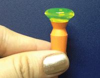 | |
| Fig. 1. Fill the lens excessively with non-preserved saline. |
The patient must tuck their chin into their chest and look down with their nose pointed to the floor. While this may sound strange to most patients, there is a legitimate reason for this: the liquid that fills the scleral lens needs to remain in the bowl.
Filling the lens excessively with non-preserved saline will help prevent insertion bubbles in the case of excessive eye movement, blinking and other errors (Figure 1). As some of the fluid will undoubtedly be spilled during the insertion process, excessively filling the lens bowl ensures there will be enough liquid left to still yield an appropriate fit.
3. DON’T let go of the insertion device first!
In one of the more common “rookie mistakes,” patients will hold their eyelids open, insert the lens, remove their insertion device (e.g., plunger, fingers, plastic ring, etc.) and then release their eyelids.
Fortunately, this mistake is also one of the easiest to remedy. After the lens is inserted on the eye, inform patients to release their eyelids first, and then release the insertion device. Releasing the eyelids first allows the lens to become trapped under the lids and onto the eye.
4. DO inform your patients of several ways to insert the lens.
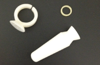
During the insertion and removal training, be sure to thoroughly explain and demonstrate several insertion techniques (Figure 2). For instance, the large plunger may work well for some patients, but many others will find that a scleral ring/orthodontic band/O-ring offers a greater amount of control.
Using these devices allows the lens to rest on one finger, freeing the others for lid control. This is particularly useful for patients who have previously worn contact lenses, as using one finger to insert the lens may feel more natural.
Additionally, training your patient on how to insert the lens using a tripod (three-finger) method may be useful for those who do not want to rely on devices to insert the lens. Giving patients a variety of insertion techniques allows them to trial several methods at home to determine which they personally find most comfortable. Simply offering patients a number of insertion options can be a huge factor in their success with scleral lenses.
5. DON’T send your patients home without resources!
After the lesson on insertion and removal training, send patients home with multiple resources (e.g., information on scleral lenses, insertion and removal handouts, lens care information, etc.). For example, I have each patient watch a 10-minute video from sclerallens.org on how to properly care for scleral lenses.
 |
|
|
Fig. 2. There are a number of scleral insertion tools and techniques.
|
Additionally, I always give them a step-by-step guide detailing the process of scleral lens wear (e.g., place the lens on the insertion device, then fill up the lens with non-preserved saline until it is almost overflowing).
If a patient forgets something, they can quickly refer to the provided guide. A quick-reference ‘scleral lens tip sheet,’ (e.g., apply make-up after inserting your lenses, wash your hands with soap that does not contain moisturizers, etc.) can also be a valuable asset for new scleral lens wearers.
6. DON’T assume a lens bubble will dissipate on its own.
After inserting a scleral lens, immediately check for bubbles at the slit-lamp. If you see any, the lens must be removed and reinserted. The presence of bubbles is typically due to an error during insertion.
Many practitioners think that as the lens settles, the bubble will move and dissipate on its own. This is not the case! The bubble will remain in the tear chamber until the lens is removed and correctly reinserted, and will greatly affect your evaluation of the fit and the over-refraction.
7. DO perform a tear exchange test at the follow-up visits.
At any of the follow-up visits, dab a copious amount of sodium fluorescein on the lens surface. Then, approximately 10 minutes later, check to see if any of the sodium fluorescein can be detected underneath the lens. This can help determine whether or not any tear exchange is occurring throughout the day.
If patients complain of red eyes, discomfort, blurred vision or burning upon lens removal, it could be because the lens edge is too tight and is not allowing any tear exchange. This is one of the most common problems seen with scleral lenses. One solution may be to flatten the edge of the scleral lens, which creates greater alignment with the conjunctiva, and allows adequate tear exchange.
8. DON’T forget to ask the patient questions at every follow-up visit.
At a patient’s first follow-up visit, lens care and insertion/removal should be detailed. Ask what solution they are using to clean, as well as what they are using to fill the lens. Additionally, ask about insertion and removal to see if they are more proficient in their technique.
Also ask about quality of vision and comfort. For example, if a patient complains of blurred vision and discomfort upon insertion that progresses as the day goes on, it is likely due to the presence of a bubble. Explaining the need to reinsert the lenses can help the patient to understand why they were having a problem in the first place, and offering a solution will give them more confidence in the product.
9. DON’T “set patients free” after their one-week follow-up.
Scleral lens patients need to be monitored more closely than conventional soft contact lens wearers. This is because problems such as lens fogging and tight lens syndrome can arise in scleral lens wearers. If a patient is doing well at their one-week follow-up visit, it would be advisable to see them in about one month, and then three months after that.
At each visit, check the lens surface for scratches and build up, then check their tear chamber for clouding before adding sodium fluorescein. Remove the lenses and check their cornea for staining, neovascularization and edema. Regularly checking for complications will help detect issues sooner, and give the patient the perception that their lenses are truly a custom product.
10. DO develop a contract for specialty lens patients.
Many insurance policies will not reimburse properly for a scleral lens fitting or for the lenses themselves. In such cases, I would advise you to create a scleral lens contract. Be sure to highlight the costs associated with both the fitting and the lenses.
Also, it is important to include a description of what the fitting fee covers (e.g., one-hour diagnostic fitting, one-hour dispense, etc.). The contract should include a section detailing what happens if the lenses don't work for any reason; does the patient get any sort of refund?
For example, I keep the entire fitting fee, but I will refund the patient the cost of the lenses—minus shipping and restocking fees. When a contract is written, read and signed, both parties know exactly what to expect.
Fitting scleral lenses can be difficult for both patients and practitioners.These 10 simple tips can help simplify the process and ensure scleral lens success.
1. Nichols, J. Annual contact lens report. Contact Lens Spectrum. Jan 2012.
2. Visser ES, et al. Modern scleral lenses part I. Clinical features. Eye and Contact Lens. 2007;33:13-20.

