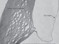
This novel layer of the cornea was identified last summer by Harminder S. Dua, MD, PhD.
“The layer is quite controversial,” says Joseph Shovlin, OD. “If it really exists, it’s probably been described by Binder decades ago in a publication where he noted some post-stromal changes next to Descemet’s membrane.”
| |
|
Dua's layer. Photo: Harminder S. Dua, MD, PhD
|
The contentious discovery is a thin (10um to 15um), acellular layer of the cornea sandwiched between the stroma and Descemet’s membrane.
Dua and colleagues believe that the collagen core of the trabecular meshwork is an extension of the PDL. In a study published in the February 16 online version of The British Journal of Ophthalmology, the team of researchers studied both the PDL and TM of 19 human donor eyes to determine the relation of the peripheral part of the PDL to the origin of the TM.1
Light and electron microscopy was used to examine the tissue structure of each donor eye, and immune-histology was examined for four matricellular proteins, five types of collagens and CD34.
The electron microscopy revealed that strands of collagen originating in the periphery of the PDL on the anterior surface of Descemet’s membrane divided and then continued as the beams of the TM. Collagens present in the PDL were found to extend into the TM. The researchers also observed matricellular proteins predominantly in the TM, with only laminin extending into the PDL.
The authors of the study believe that these findings may aid future research into both the TM and glaucoma by helping to better understand aqueous outflow mechanisms and potential pharmacologic interventions.
1. Dua HS, Faraj LA, Branch MJ, et al. The collagen matrix of the human trabecular meshwork is an extension of the novel pre-Descemet’s layer (Dua’s layer). Br J Ophthalmol. 2014 Feb 16. [Epub ahead of print]

