Complications of the cornea, such as microbial infections and allergic reactions, can present in a number of ways, and symptoms can vary from simple discomfort to vision loss in extreme cases. Due to the complexity of these presentations and the multitude of potential causes for each, it’s crucial to assess and diagnose the myriad complications in a timely manner in order to properly and promptly treat your patients. This photo gallery can help guide the way you handle these challenging conditions.
Subconjunctival Hemorrhage
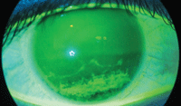
I like to tell my patients that everybody is allowed one subconjunctival hemorrhage in their lives. While these hemorrhages look terrible, they are actually benign. But when a patient presents with a subconjunctival hemorrhage, it could be an indication of a deeper problem. So, it’s important to ask the patient if they are taking blood thinners or have a blood disorder, such as leukemia.
If the patient has chronic subconjunctival hemorrhages or bruises elsewhere on their body, they should be worked up for blood dyscrasia. Patients are often beside themselves with worry over these hemorrhages, but the ocular presentation will not result in any loss of vision.
 |
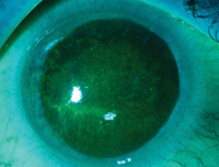
|
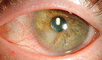
Significant subconjunctival hemorrhage in a patient on blood thinners.
|
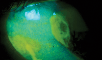
Corneal Staining
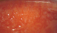
These photos demonstrate just a few of the many different types of staining we see on the eye in clinical practice. The key to these photos is that the pattern of the staining indicates the specific pathology and illuminates the diagnosis. In the first case, there’s a visible line along the eyelids, indicative of lagophthalmos, while the patient in the second photo exhibits overall diffuse staining, which indicates a toxic event present in the eye.
 |

|
|
Staining from nocturnal lagophthalmos.
|
Diffuse staining from toxic keratopathy.
|
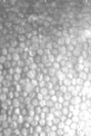
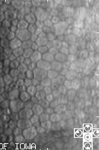
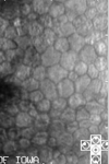
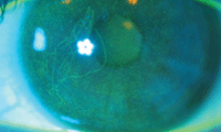
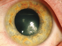
Endothelial Morphology
These photos display polymegethism (cells enlarging to take up the space of adjacent cells that died) and pleomorphism (cells that have changed their shape and are no longer hexagonal) changes as endothelial damage progresses. We are born with about 3500 endothelial cells per mm2. Throughout the course of our lives, natural aging will cause some of these cells to die and drop off. In a normal, healthy adult, there are approximately 2500 endothelial cells per mm2.
 |
|
|
|
|
Normal endothelial morphology.
|
Mild endothelial morphology.
|
Moderate endothelial morphology.
|
Severe endothelial morphology.
|
When we observe a drop to fewer than 800 cells per mm2, the cornea becomes pathologic and experiences edema.
As doctors, even though we can’t count the number of cells present at the slit lamp, we must remember the importance of the endothelium and the overall functioning of entire eye. It’s possible that we may put a contact lens on the eye that stresses the endothelium, accelerating the death of cells. Which particular contact lens we fit someone in at age 20 may have an impact on that person at age 80.
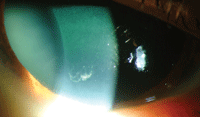
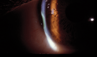
Microbial Keratitis
Microbial keratitis can be treated with broad-spectrum antibiotics. If the infection presents in a central location and threatens sight, a culture may need to be done. If the borders of the presentation are not round, or have an otherwise unusual presentation, we usually recommend culturing to determine the organism.
|
|
|
Staining in Psuedomonas keratitis.
|
Paracentral Psuedomonas keratitis.
|
Unfortunately, it is usually not practical to do a culture prior to initiating therapy, so first start with broad-spectrum antibiotic. If the keratitis doesn’t respond, culture the organism and adjust as needed. Often, doctors are afraid that if they begin with a course of antibiotics, it’ll throw off the culture results, but it’s important to keep in mind that most cultures are not going to come up positive, regardless of what we do.
|
|
|
Stromal necrosis in a patient with microbial keratitis.
|
Stromal staining over necrosis in
microbial keratitis.
|
Timing of the culture will not generate a positive or negative reading—if you culture later on, and the organism is still active in the area; the culture will come up positive, regardless of antibiotic use.
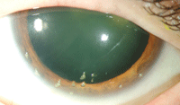
Giant Papillary Conjunctivitis
There are two forms of GPC: allergic and mechanical. Commonly, we think of GPC as being caused by an allergy—our body’s way of reacting to allergens by creating the papilla to get allergens out of eye.
With the advent of SiHy lenses, we’ve actually started seeing mechanical GPC from the modulus of the lens causing rubbing on the tarsal plate, which causes focal GPC. This form is not allergic and cannot be treated as such. The only way to treat patients presenting with mechanical GPC is to remove the lens entirely.
|
|
Diffuse allergic GPC in a lens wearer.
|
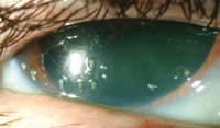
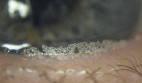
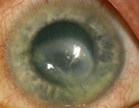
Filamentary Keratopathy
Filamentary keratopathy refers to mucin strands attached to corneal epithelial cells. This condition occurs in patients presenting with severe dry eye. Frequently, filamentary keratopathy will occur in the area of exposure.
Symptoms of the condition can range from foreign body sensation to significant pain. The common treatment for patients with filamentary keratopathy is a bandage contact lens, but more severe cases will require tarsorraphy.
|
|
|
|
Filamentary keratopathy presenting in the inferior cornea.
|
Multiple filaments in a patient with severe dry eye.
|
Filamentary keratopathy may be treated with a bandage contact lens.
|
FB Tracking
This particular photo is of a patient who got plaster in his eye. So, in this particular scenario we faced two things: toxic keratopathy from the lye in the plaster, and hunk of plaster lodged under the eyelid, causing foreign body tracking.
Any time a patient presents with foreign body tracking, you must evert the upper eyelid to see what’s underneath. In addition to debris in the eye, this patient also had a toxic reaction, so we had to irrigate his fornices to prevent chemical burn.
Scenarios like this emphasize the importance of considering and understanding the causative agent specific to each case as well as the circumstances of the injury, as treatments may vary.
|
|
Foreign body tracking from debris under upper eyelid.
|
Dellen
This is an example of what can happen to a patient who presents with chronic dry eye. When the cornea is exposed to the environment and is not properly lubricated, the stroma will start to thin out. This thinning is referred to as dellen.
In severe cases, it can perforate. It is not infectious and does not have to be treated as such. As indicated by the staining photo, there is no break in the epithelium into the stroma, and while there is surface dryness, the area over the dellen remains intact.
|

|
|
|
Stromal thinning from chronic exposure in a dellen patient.
|
Staining of the conjunctiva and cornea in a dellen patient.
|
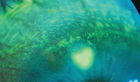
No staining of the dellen bed and no stromal
fluorescein uptake.
|
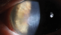
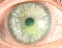
Corneal Edema and Folds Under Scar
This is a result of severe dry eye; the long-term nature and severity has caused not merely scarring but also a complete corneal breakdown. The resulting dysfunction of the endothelium causes corneal folds.
This photograph illustrates the importance of getting any severe dry eye condition under control, as it can completely destroy the cornea if left untreated.
|
|
Corneal edema and folds beneath scarring.
|
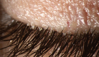
Anterior Blepharitis/Demodex Infestation
Demodex is a parasitic arachnid commonly found in the environment. Of the many species of Demodex, only two are present in humans, and they are not the same that live on pets. It is unknown how much the organism influences the course of pathology on the ocular surface; however, when there is significant blepharitis present, often there is also a significant Demodex population.
Symptoms of Demodex are collarettes along the base of lashes, itching, poor tear film quality and ocular surface hyperemia.
|
|
Collarettes around the base of the lashes is a predictable sign of Demodex.
|
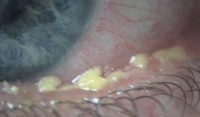
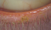
Blepharitis
Meibum is a vital component of the lipid layer of the tear film, which prevents evaporation. Inconsistency in meibum secretion due to blepharitis alters tear functionality, causing chronic dryness. Telangiectasia is a sign indicating chronic inflammation of the eyelid margin.
This indicates that the blepharitic state has been there for a significant amount of time, and the margin is undergoing significant changes. If telangiectasia is observed, it is a clear sign of the chronicity of the blepharitis.
|
|
|
|
Thick meibum secretions in posterior blepharitis.
|
Saponification of the lipid layer of the tear film in a patient with blepharitis.
|
Telangiectasia in a patient with chronic blepharitis.
|
Acanthamoeba
It’s important to observe a variety of Acanthamoeba presentations, because the condition itself often masquerades. Frequently, Acanthamoeba is not the first thing doctors think of; because of the condition’s subtle presentations, sometimes practitioners do not consider the condition at all.
|
|
|
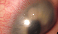
Dense epitheliopathy and a
pseudodendrite on initial
presentation of Acanthamoeba.
|
Acanthamoeba dine on bacteria. Suspect the organism in ulcerative looking lesions that do not resolve with antibiotics.
|
This is problematic if it delays diagnosis and the prompt initiation of therapy, further exacerbating the situation.
|
|
|
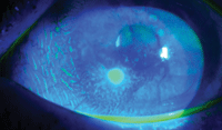
The initial signs of Acanthamoeba may be subtle.
Most present with mild epitheliopathy. |
Classic Acanthamoeba rings do not appear until
later in
the infection. |
Dr. Sindt is the Associate Clinical Editor of Review of Cornea and Contact Lenses, and of Review of Optometry. She is also the Director, Contact Lens Service and an associate professor of clinical ophthalmology and visual sciences at the University of Iowa.

