The early history of contact lenses can also double as a history of treatment for irregular corneas. In a 1827 publication of the journal Light, Sir John Herschel suggested that a lens could be ground to the surface shape of the cornea. “Should any very bad cases of irregular cornea be found, it is worthy of consideration whether at least a temporary distinct vision could not be procured by applying in contact with the surface of the eye some transparent animal jelly contained in a spherical capsule of glass, or whether an actual mold of the cornea might not be taken and impressed on some transparent medium,” he wrote.
The first actual contact lenses, produced by Adolf Fick and Edouard Kalt, were designed as blown glass shells and introduced in 1887. The goal was to improve vision in keratoconus patients and to protect the corneas from the ravages of trachoma.
In 1888, August Muller produced the first lens with refractive power. Other major developments in contact lens technology include William Feinbloom’s use of PMMA for scleral lenses in 1936, the first commercially successful PMMA corneal lenses by Kevin Tuohy in 1949, George Butterfield’s first corneal lenses in 1950, Otto Wichterle’s development of HEMA materials in the 1960s and the development of gas-permeable rigid lenses in the late 1970s.
Undoubtedly, the most satisfying part of any contact lens practice is the ability to restore that most precious sense—sight. Each new breakthrough has given us the ability to help those with visually compromising corneal irregularity and distortion so they function normally.
Treating Keratoconus
Almost any condition that causes an irregular corneal surface in the absence of any active infectious process can be helped optically and in many instances therapeutically by the application of some type of contact lens.
The earliest use of contacts was for the treatment of keratoconus—a non-inflammatory, usually bilateral, progressive asymmetric ectasia of the cornea resulting in poor vision due to the distortion produced by the disease. It was thought early on that applying a lens, which gently touched the apex of the cone, would retard the progression of the disease and improve visual function. Later studies showed that this use could cause scarring of the cone.
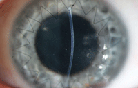
PK with sutures still in place.
There have been many lens options to treat keratoconus over the years: glass, PMMA scleral lenses, RGP corneal lenses, soft toric lenses, specially designed soft lenses, hybrids and high dk scleral lenses.
• GP. These remain the most prescribed modality for this condition: spherical designs and aspherics for early cones, and specialty designs (such as Soper Cone, McGuire Cone and various Rose Cone lenses) for more advanced cases.
• Disposables. Piggyback techniques, such as using a disposable soft lens with a GP lens, can provide added comfort for the patient who cannot tolerate a rigid lens due to sensitivity issues. There are a number of soft lenses designed to hold the rigid lens in place, but in many cases a standard disposable lens will work as well.
• Specialty Lenses. Another option to improve comfort is the use of specialty keratoconus soft lenses such as the NovaKone (Alden Optical), KeraSoft IC (UltraVision) or HydroKone (Visionary Optics). These lenses can be made with steep base curves (4.1mm) and are thicker than average to help mask the corneal irregularity. The sphere or spherocylinder power is placed on the front surface. These are the most advantageous when used for spherical, more central cones; decentered or sagging cones see less success with these lenses.
• Hybrids. These lenses have long been used to treat keratoconus—with fairly poor results due to the limited parameter range. With the development of SynergEyes hybrids, we now have a wide variety of design options, central curvatures and variable peripheral curves. Early cones are best fitted with the standard A series or Duette lens, while more advanced cones are better suited to the KC series or the Clear Kone design.
 The Use of Bandage Lenses
Epithelial, stromal and endothelial dystrophies can cause corneal surface irregularity, which can be treated with therapeutic contact lenses and concomitant drug therapy. This treatment plan will increase vision, relieve pain and decrease photophobia that often accompanies these conditions.
Recurrent corneal erosions will heal faster with the use of a bandage lens; extended use may reduce the incidence of recurrence. The lens reduces pain and the foreign body sensation that many patients describe, along with reducing glare and photophobia. Concomitant antibiotic therapy and lubricating drops reduce the chance of secondary infections and keep the lens moist.
Soft bandage lenses can also ease the foreign body sensation and photophobia caused by epithelial basement membrane disease, Reis-Bucklers’ corneal dystrophy and Meesmann dystrophy. They can also be used to reduce recurrent erosions, foreign body sensation, and pain and light sensitivity for stromal dystrophies such as granular dystrophy and lattice dystrophy. Large bullae associated with Fuchs’ endothelial dystrophy or aphakia can cause severe pain and blurred vision, which can be greatly helped with bandage lenses.
Post-Surgical Lens Applications
• RK. Radial keratotomy (RK) was a popular treatment in the 1980s, but is rarely used today due to wound healing problems and post-surgery side effects that may leave patients hyperopic and with distorted corneas. Many of RK patients can’t or won’t wear glasses, and are instead returning for contact lens correction. However, the oblate nature of these corneas makes them a difficult fit for standard GP lenses. Soft lenses may promote neovascularization into the RK cuts, and due to the vaulting affect over the flattened central cornea, may not give the patient good visual acuity.
Standard GP designs center around the steepest area of the cornea, which has moved post-surgery. Instead, large diameter intralimbal designs, or even semi- and mini-scleral lenses, may be needed to attain good lens centration.
To start a fitting, use the flattest K reading prior to the RK procedure as your base curve. The final lens Rx will be similar to the preoperative Rx since you are vaulting the flatter central area. Keep in mind that you are looking for peripheral alignment. If the original K readings are not available, gauge the curvature of the cornea through topography—4mm lateral to the center of the cornea is where you want to start your trial fitting. Highly oblate corneas may need to be fitted with reverse geometry lenses for better alignment and more centration.
• PRK, LASIK. PRK and LASIK are generally highly successful refractive procedures, but the small percentage of failures can be catastrophic to the patient, resulting in blurred or distorted vision, monocular diplopia, photophobia or corneal ectasia. Decentered ablations can cause high degrees of irregular astigmatism. Standard large diameter lenses are needed on these usually oblate corneas; intralimbal designs with 11mm to 13mm diameters work well. Semi- and mini-scleral lens from 13mm to 18mm can provide even better centration. Patients with significant dioptric differences between the central cornea and periphery who find that bubbles may be trapped over the central cornea in standard lenses may do well with the reverse geometry designs.
Hybrids can also be used if comfort with GP lenses is an issue. Patients with ectasia will benefit from the SynergEyes KC series; more oblate corneas do well with the reverse geometry design of the SynergEyes PC lens. The ClearKone also seems to work well when other designs fail. The ultimate goal is to have a well centered lens that gives good endpoint vision with no central bubbles and no vessel impingement of conjunctival vessels along with good movement and tear exchange.
• PK, DALK. Penetrating keratoplasty (PK) or deep anterior lamellar keratoplasty (DALK) is usually indicated for advanced keratoconus that cannot be corrected with contacts, central corneal scars secondary to trauma or severe corneal disease such as herpes kerititis, corneal ulcers or dystrophic conditions. Possible post-surgery side effects include corneal distortion or high degrees of regular or irregular astigmatism. The fitting process can start as early as three months after surgery, even with the sutures in place. If a patient’s sutures have been removed to limit the possibility of blood vessel growth along the suture lines and across the host donor junction, soft spherical or toric lenses are recommended. Deep vessel invasion of the graft could cause clouding, edema or graft rejection.
Rigid lenses of 11mm and larger can vault the host donor junction and usually give good optical and physiological results. The fit will have to be closely monitored for six to nine months postoperatively as the healing process may cause reshaping of the cornea.
When fitting a lens early in the healing process, the donor button may be depressed in relation to the host cornea. Standard lens designs may vault the central cornea and large bubbles may be trapped centrally. In these cases, reverse geometry lenses may give a better response. As the button steepens with time, a change to a standard base curve lens may be necessary. Be sure to carefully explain the course of the healing process to the patient before starting the fitting process so as to avoid any misunderstandings. The patient also needs to know that if and when the sutures are removed, the fitting process may need to be repeated since the cornea can change significantly.
Scleral lenses (from 18mm to 24mm) are having a significant impact in cases where standard size lenses fail. These larger diameter lenses are fitted so that a fluid reservoir is maintained under the lens, which will mask any corneal irregularity. Here, trial lens fittings are essential to get the best results. Use these lenses for severe keratoconus, post-surgical cases where smaller lenses have failed and to aid patients with severe ocular surface disease.
As we continue to address complications that stem from irregular corneas and their subsequent treatment, we are fortunate to see the development of new technology to better fit the patient.
Dr. Weiner is a graduate of the Pennsylvania College of Optometry. He has served as an assistant professor of ophthalmology at the University of Maryland Medical School and currently serves as ancillary faculty at the Wilmer Eye Clinic. He is the past chair of the American Optometric Association’s Cornea and Contact Lens section and is a fellow of the American Academy of Optometry.

