 Nodular corneal degenerations can lead to both decreased vision and ocular discomfort, and the range of findings can vary greatly.1 It is more commonly bilateral, but a significant percentage of patients have unilateral findings.1 Nodular changes occur predominantly in middle-aged women and may be associated with chronic ocular surface inflammation and/or irritation.1 Patients with nodular corneal degenerations most often seek surgical treatment for visual symptoms more so than irritation.1
Given that the vision issues that lead patients to seek surgical intervention are caused mainly by irregular astigmatism, it would seem that gas-permeable (GP) lenses would be an option for treating nodular degenerations. However, one of the problems with using GPs for correcting irregular astigmatism in these patients is the contact between the rigid lens and the corneal nodules. In most cases, patients with nodular corneal degenerations will not tolerate standard GP lenses. In these cases, opting for methods of correcting their vision with non-standard approaches can be very effective.
In this column, I will discuss the first of two such patients and the two different approaches that led them both to successful lens wear.
A Case Study
JF, a 33-year-old white female, visited my office in September 2005 at the request of her corneal specialist for replacement of a bandage soft contact lens in her left eye. She had a long-standing history of Salzmann’s nodular corneal degeneration (SNCD) in both eyes, and had a corneal transplant in the right eye a few years prior.
The transplant procedure in the right eye had not gone as expected and the vision in that eye was very poor at 20/400. The vision in her left eye was not much better (best-corrected visual acuity was 20/200), but due to the poor outcome of the right eye, she was not interested in a corneal graft O.S.

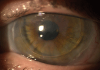
1. Salzmann’s nodular corneal degeneration O.S. with piggyback lenses in place.
She continuously used bandage soft lenses in both eyes to alleviate symptoms of chronic irritation; the bandage lenses were removed and replaced every two months in an off-label manner. The bandage lenses were well-tolerated, and JF presented with no new complaints. As this was my first visit with her, I had no knowledge of her history beyond what her chart indicated. I evaluated her corneas and her vision and asked her how she was functioning.
She reported that, in the last five years, her vision had deteriorated to the point where she was unable to drive or hold a job, which was why she had the transplant on the right eye. Unfortunately, the corneal transplant had not been effective to date with episodes of rejection, infection and dehiscence with repair.
At the time of her presentation, her right eye had significant scarring of the cornea due to prior infection, as well as a tube shunt for complicated glaucoma. There was little to do for this eye at the moment.
Upon examination of her left eye, there were numerous nodules covering the entire corneal surface, creating haze and irregularity (figure 1). Refraction over the top of the bandage contact lens provided no improvement. I asked JF if anyone had ever suggested using a GP lens in her left eye to try to improve her vision. She said that she had not, but was willing to try.
After consulting with her corneal specialist and getting approval from him, I placed an average base curve lens on her left eye over the soft bandage lens. Her initial impression of the comfort was as positive as could be expected, but the visual acuity was overwhelming. With the trial lens on, her vision was 20/50. With a small over-refraction her vision improved to 20/25-1. The lens was essentially a lid control fit over the soft bandage lens.

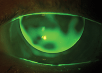
2. Fluorescein pattern of left eye with Salzmann’s nodular corneal degeneration and piggyback contact lenses in place.
Fluorescein pattern evaluation was done using high molecular weight fluorescein, which clearly demonstrated the significant corneal irregularity caused by the SCND (figure 2).
The final lens was ordered with the following parameters in a thin design: 7.85 O.S./-3.50D/9.5mm. Because the irregularities do not occur across a common axis, a thin lens design was very effective at covering the irregularities and maintaining good comfort. Because the soft lens was used under the GP, the corneal surface did not suffer breakdown or irritation from contact with the GP lens.
The patient continued to use the same soft lenses as she had previously worn, and vision continued to be stable in the 20/25 to 20/30 range. She applied and removed the GP lens daily, but left the soft lens in for two months as she had always done. JF continues to use this modality of correction more than six years later, and vision and comfort are maintained as the same levels as at the initial fit.
Further Discussion
Nodular corneal degenerations can be difficult to manage. Surgical intervention often can be helpful, but carry significant risks in eyes with chronic inflammation, and nodules are shown to recur in eyes after surgical therapy.2-4
Penetrating keratoplasty can be an option in severe cases, though it carries all the standard risks, in addition to likeliness of recurrence. Instead, phototherapeutic keratectomy (PTK) is the procedure of choice in recent years—it carries less risk than keratoplasty while achieving comparable results.5
Keep in mind that recurrence is still a risk when performing PTK for nodular corneal degenerations.6 Practitioners can apply 0.02% mitomycin C to the post-keratectomy corneal surface with a cellulose sponge for 30 seconds to 120 seconds to reduce both the risk of nodular recurrence and haze.6,7
When effective at correcting vision and well tolerated, GP lenses can be considered as a treatment for patients with irregular astigmatism from SCND. Because of potential problems with GP lens and corneal interaction, piggybacking or scleral lenses are some options that can minimize or avoid contact and should be considered as first choices. In the next column, we’ll discuss a similar case managed with scleral lenses.
1. Farjo AA, Halperin GI, Syed N, Sutphin JE, Wagoner MD. Salzmann’s nodular corneal degeneration clinical characteristics and surgical outcomes. Cornea. 2006 Jan;25(1):11-5.
2. Sinha R, Chhabra MS, Vajpayee RB, et al. Recurrent Salzmann’s nodular degeneration: report of two case and review of literature. Indian J Ophthalmol. 2006 Sep;54(3):201-2.
3. Severin M, Kirchof B. Recurrent Salzmann’s corneal degeneration. Graefes Arch Clin Exp Ophthalmol. 1990;228(2):101-4.
4. Yoon KC, Park YG. Recurrent Salzmann’s nodular degeneration. Jpn J Ophthalmol. 2003 Jul-Aug;47(4):401-4.
5. Sharma N, Prackash G, Titlyal JS, Vajpayee RB. Comparison of automated lamellar keratoplasty and phototherapeutic keratectomy for Salzmann nodular degeneration. Eye Contact Lens. 2012 Mar;38(2):109-11.
6. Marcon AS, Rapuano CJ. Excimer laser phototherapeutic keratectomy retreatment of anterior basement membrane dystrophy and Salzmann’s nodular degeneration with topical mitomycin C. Cornea. 2002 Nov;21(8):828-30.
7. Khaireddin R, Katz T, Baile RB, et al. Superficial keratectomy, PTK, and mitomycin C as a combined treatment option for Salzmann’s nodular degeneration: a follow-up of eight eyes. Graefes Arch Clin Exp Ophthalmol. 2011 Aug;249(8):1211-5.


